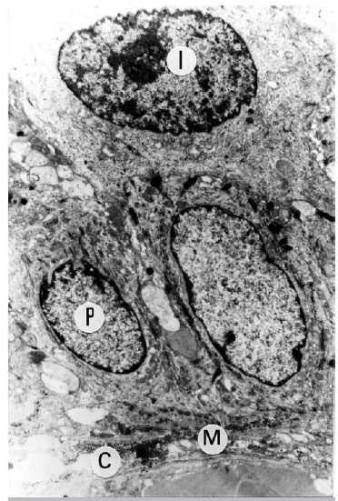
Figure 1. TEM of case No. 19, image showing ultrastructural appearance of neovaginal mucosal lining, similar to a normal mucosal lining, presenting chorion (C), basal membrane (M) and, in this epithelium, deep (P) and intermediate (I) layer cells are noted. Cytoplasmic and cell organelle characteristics are similar to those of a normal epithelium.
Image size increased 7,300 fold.