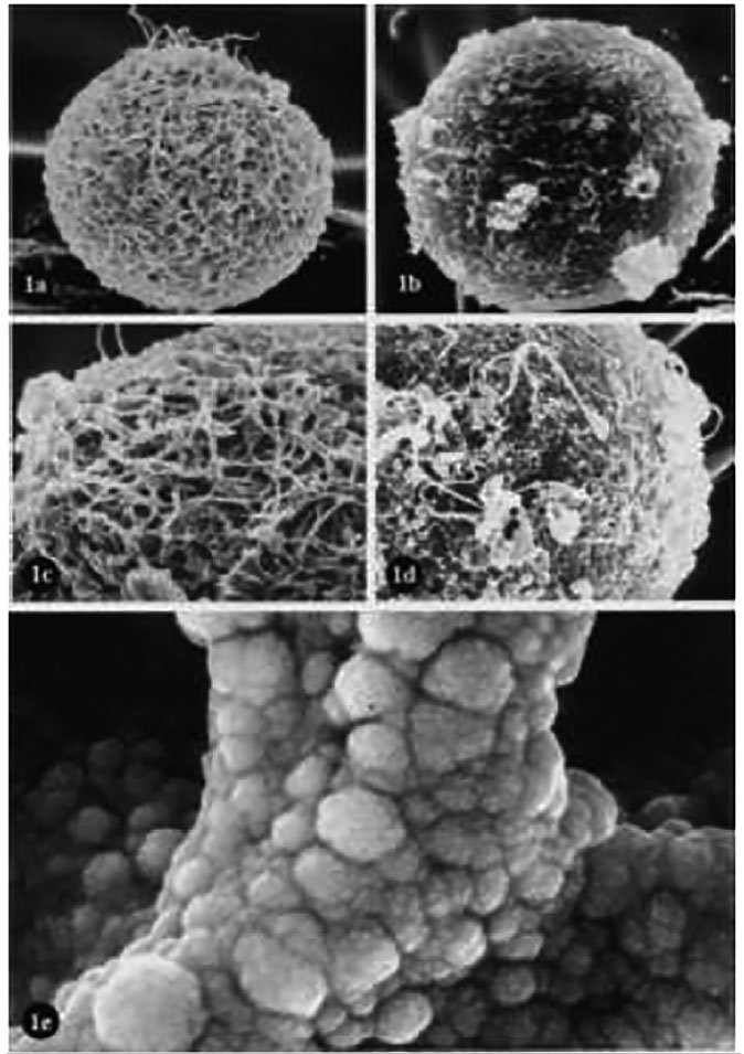
Figure 2. Conventional SEM. (a): Unfertilized human oocyte. Porousnet appearance of ZP (X 2,000). (b): Unfertilized human oocyte. Compact and smooth surfaced ZP (X 2,000). (c): Higher magnification of (a). The spongy ZP structure is evident (X 4,000). (d): Higher magnification of (b). It shows a dense and compact ZP structure (X 4,000). (e): Unfertilized mouse oocyte with very high magnification showing a branch of the spongy structure of the ZP (X 350,000). Familiari et al. (2006) with permission.